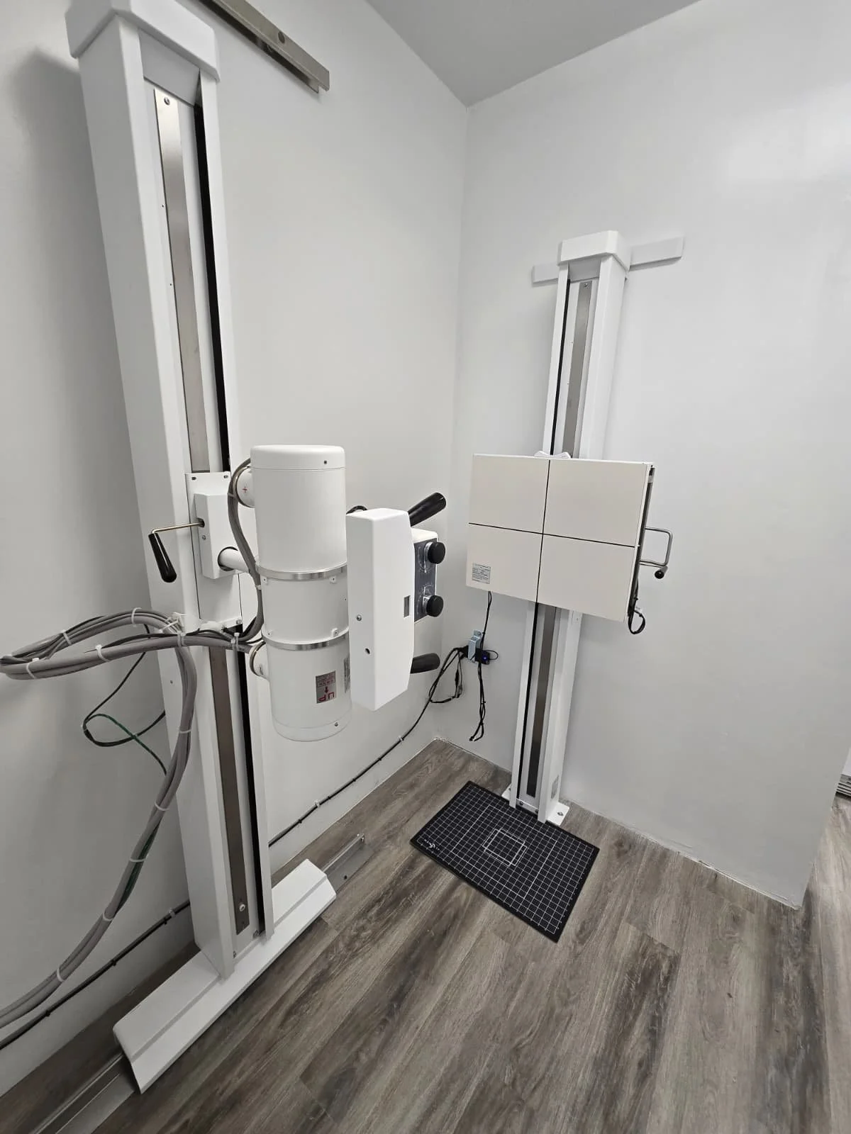
X-Rays
X-rays use invisible beams of electromagnetic energy to produce images of internal tissues, bones, and organs on film or digital media. Standard X-rays are performed for many reasons, including diagnosing tumors or bone injuries.
X-rays play a vital role in chiropractic care by providing detailed images of a patient’s spine and surrounding structures. These images help chiropractors accurately diagnose underlying conditions, identify spinal misalignments, and detect abnormalities such as degenerative disc disease, scoliosis, arthritis, fractures, or congenital defects. With this information, chiropractors can create a safe and effective treatment plan tailored to the patient’s specific needs.
By visualizing the position and condition of the vertebrae and discs, chiropractors can determine if it is safe to proceed with manual adjustments or if alternative care is necessary. X-rays also allow practitioners to identify any red flags that may require referral to a different healthcare provider, such as signs of infection, cancer, or severe trauma.
While not all patients require X-rays, they are particularly useful for those with a history of trauma, unexplained pain, or signs of spinal instability. X-rays enhance diagnostic accuracy, reduce guesswork, and support evidence-based care, ultimately improving patient outcomes. X-rays are a powerful tool that supports the chiropractor in delivering safe, personalized, and effective spinal healthcare at prices accessible to any budget.
In cases of chronic pain or post-injury rehabilitation, X-rays help monitor
progress and assess whether the spine is responding to treatment. Structural chiropractic techniques, like Chiropractic BioPhysics® (CBP®), use precise spinal measurements from X-rays to guide corrective care aimed at restoring normal spinal curves and posture.


